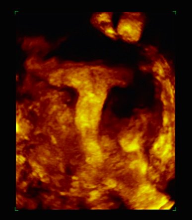
- This technology is used in all IVI centers, allowing the comprehensive assessment of the uterus, in a simple, fast and detailed way
- The “T-uteri”, which is characterized by a narrow and tubular endometrial cavity, lead to a higher frequency of worse reproductive and obstetric results
DENVER, OCTOBER 8th, 2018
The growing importance in the investigation of the denominated “uterine factor” in Human Reproduction is a challenge to improve pregnancy rates. This is the starting point of a study entitled “T-shaped uterine cavity morphology as assessed by three-dimensional ultrasound (3D US) may be associated with lower implantation rates and higher clinical rates following frozen embryo transfer”, which IVI will present at the 74th edition of the American Society for Reproductive Medicine Congress (ASRM) in Denver (Colorado) from October 6th to 10th.
In this research, IVI analyzes and evaluates, using 3D ultrasound the mullerian malformation known as “uterus in T”, demonstrating the usefulness of this technology to help improve the reproductive prognosis of patients suffering from this anomaly.
“This diagnostic method, which we firmly believe in at IVI – both in the research and clinical aspects – is available in all our clinics. 3D ultrasound allows the comprehensive evaluation of the uterus, in a simple, fast and detailed way. Therefore, obtaining coronal uterine planes which allow us to assess the morphology of the endometrial cavity directly, this factor provides a remarkable advance over conventional ultrasound techniques based on 2D “, explains Dr. Antonio Requena, Medical Director of IVI.
The so-called “uterus in T” (anomaly in its formation during the embryonic period that involves the characteristic narrow and tubular form of its endometrial cavity) entails a higher frequency of worse reproductive and obstetric results. There is a greater occurrence of dysmenorrhea, a higher frequency of embryonic implantation failures and repeat abortions, as well as greater frequency of premature births.
The study analyzed 651 patients the day before the scheduled transfer of embryos, assigning them a morphology of the uterine cavity. There was a trend towards less implantation and greater clinical loss for patients with a T-shaped uterus, although these findings did not reach statistical significance.
“The early diagnosis of the” uterus in T “by echo3D is a potential weapon for the improvement of reproductive and obstetric areas for women, as well as being a very useful resource in the hysteroscopic operative field. The great advantage of the portability of the eco3D systems means that all IVI operating rooms have these systems in their daily functioning, which is a useful tool to support the surgeon in the surgical remodeling of these narrowed endometrial cavities in those cases in which there is surgical indication. In addition, this technology allows us to optimize the surgical result and reduce the operative times “, adds Dr. Requena.
The studies presented by IVI at the ASRM Congress 2018 constitute a step forward in the current technological career in the evaluation of uterine pathologies for the improvement of reproductive results, with minimally invasive and highly precise methodologies.
