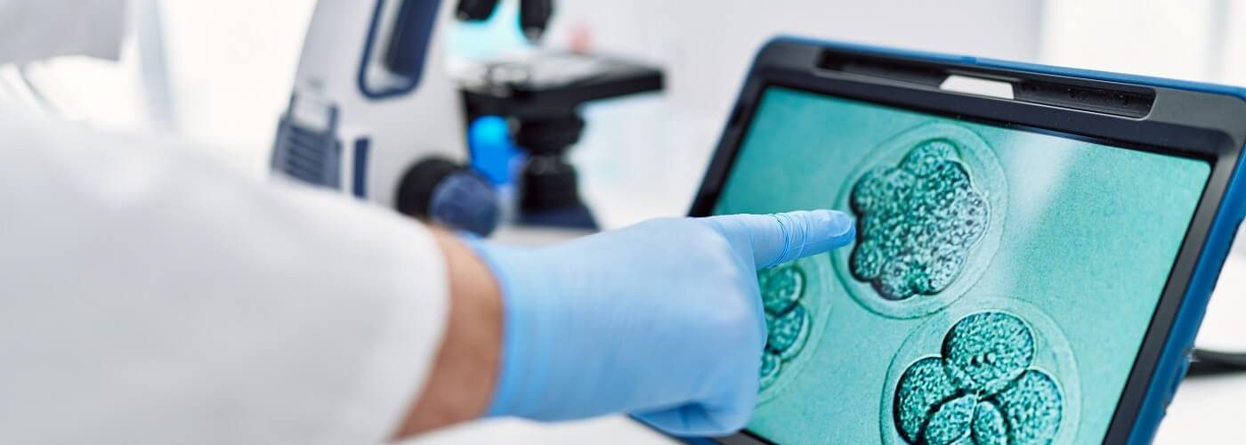
The future of medicine may lie in one of the findings presented at the 10th IVIRMA Congress, held in Malaga recently. A team of researchers led by Dr. Jacob Hanna, from the Department of Molecular Genetics at the Weizmann Institute of Science, has created artificial embryos from synthetic mouse cells. These embryos have been developed in a device with identical conditions to those in the uterus.
In this blog, we explain the details of this research. It has been presented at an event attended by more than 1500 professionals from all over the world.
Creating artificial embryos with synthetic cells
Dr. Hanna’s team has built on previous advances in their lab, such as reprogramming stem cells and returning them to their earliest stage. They also had a device capable of reproducing the action of the uterus in embryo development. This is made possible by a system of vessels in continuous movement to deliver nutrients, in the same way as the blood flow delivers nutrients to the placenta. In addition, oxygen exchange and atmospheric pressure are strictly controlled.
For this new study, the team’s goal was to grow a synthetic embryo model solely from mouse stem cells that had been cultured for years in a Petri dish, dispensing with the need to start from a fertilized egg. Before placing these cells in the ex-utero device, they divided them into 3 groups. In one of them they were left as they were and two others that were pretreated to produce extraembryonic tissue. When mixed in the device, 0.5% formed spheres that developed into an embryo-like structure. Subsequently, the researchers were able to observe the placenta and yolk sacs forming outside the embryos and the development of the synthetic model in the same way as in a natural embryo.
“When compared with natural mouse embryos, the synthetic embryos showed 95% similarity in both the shape of the internal structures and the gene expression patterns of the different cell types. The organs observed in the models gave every indication of being functional,” said Dr. Hanna.
Developing organs from artificial embryos
The result was a synthetic mouse embryo model with progenitor or specialized cells. It showed a beating heart, a brain with well-formed folds, a yolk sac, a neural tube, an intestinal tract, a placenta, and an incipient blood circulation at only eight days of development, almost half of the 20 days of gestation required for a mouse.
Now, the more realistic long-term goal is to study how stem cells form various organs in the developing embryo. This would open up new therapeutic horizons in organ transplants. Indeed, it may one day be possible to grow tissues and organs using synthetic embryo models.
As Dr. Hanna, associate professor at the Weizmann Institute of Science, explains: “The embryo is the perfect starting point for generating organs and the best 3D bioprinter, and that is the key to being able to create mechanisms that allow us to ensure that stem cells differentiate from specialized cells in the body or that they directly form entire organs. To date this has been very difficult to achieve, and the key to success has been unlocking the stem cells’ self-organizing coding potential.”
Next steps in the research
Before cells can be developed for therapeutic purposes, these stem cell transitions during embryogenesis and organogenesis must be observed. The degree of equivalence of the cells in vitro to those in vivo must also be studied. For this, it is necessary to understand their reprogramming and differentiation mechanisms.
Once we have studied and assimilated how stem cells form various organs in the developing embryo, the next challenge is to understand how the stem cells know what to do. That is, how they self-assemble into organs and find their way to their assigned places within an embryo. This system may also be useful for modelling birth and implantation defects in human embryos.
“Instead of developing a separate protocol to grow each cell type, e.g., kidney or liver cells, perhaps one day we can create a synthetic embryo-like model and then isolate the cells we need. We will not have to dictate to the emerging organs how they should develop. The actual embryo does it better,” concluded Dr. Hanna.
A breakthrough in avoiding the use of animals in experiments
The development of artificial embryos for research brings other advances for research and biotechnology. Firstly, it reduces the use of animals in experimentation. It would also avoid the technical problems that arise with the use of embryos. Even when using mice, certain experiments are currently unfeasible because they require thousands of embryos. With models derived from mouse embryonic cells, which grow in laboratory incubators by the millions, this option is virtually unlimited.
Finally, it would put an end to ethical questions about the use of animals or living beings for medical experimentation.
10th IVIRMA Congress: a landmark event
The tenth edition of the IVIRMA Congress has been held in Malaga from 20 to 22 April. It has attracted the world’s leading researchers in the sector. At this event we have presented the advances achieved in the field of Reproductive Medicine, the most innovative techniques, and the results of the latest research. In addition, it was a meeting point for sharing best practices to improve the daily results in this activity. The Congress brings together more than 1,500 specialists from 57 countries and is held every two years.


Comments are closed here.